
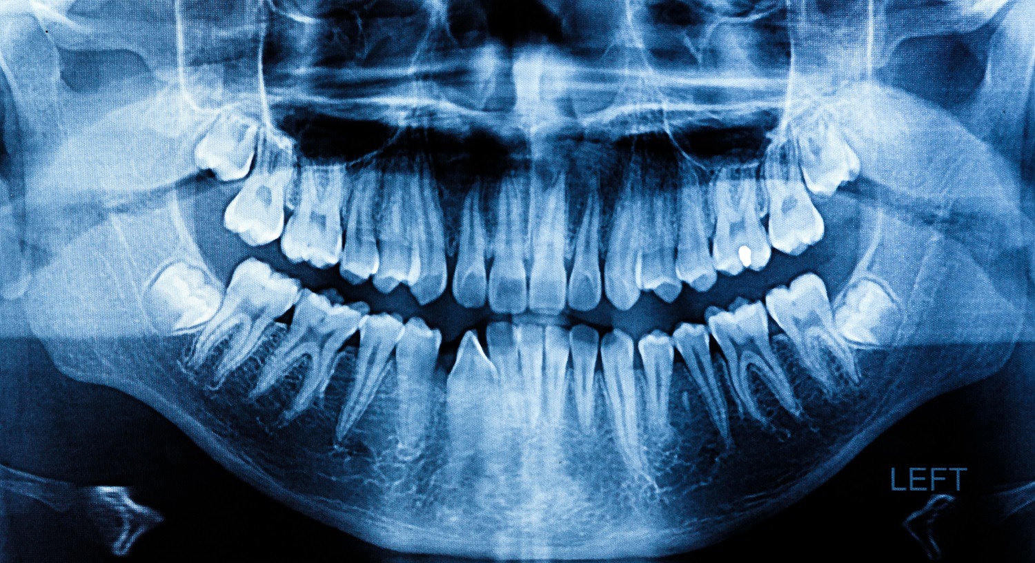
Ever wonder what the point of a dental x-ray is? Usually, it’s to check for cavities; that’s the mundane use case it experiences most often. But every once in a while, a routine scan can uncover something the naked eye never would have caught. Not a standard cavity, but something much more severe. A shadow on the bone. An irregular shape that doesn’t match the tooth next to it. That raises a real question: what does cancer look like on a dental x-ray?
The short answer is that it rarely looks dramatic. Bone destruction, odd density changes, and irregular patterns are the kinds of clues a dentist is trained to notice, long before a patient feels anything wrong.
Knowing what these signs look like can change how a person reads their own results the next time they sit in that chair.
How a Standard Dental X-Ray Can Catch More Than Cavities
Modern dental x-rays are a far cry from the thick film and metallic taste of decades past. Most offices now use digital imaging systems, which let the dentist enlarge an image on-screen and inspect small areas more closely.
It’s a huge help when spotting small, subtle changes: a faint dark patch near a root, an edge that looks worn away instead of smooth, or a spot where bone should be dense but isn’t.
Of course, a clearer image doesn’t replace a biopsy or a specialist’s opinion. It just highlights reasons a dentist may want to look closer or refer the patient for another opinion.
What Does Cancer Look Like on a Dental X-Ray?
Cancer doesn’t announce itself the way it does in the medical textbooks. There’s no glowing mass or bold outline pointing to the problem.
Instead, a dentist looks for patterns that break from what a healthy jaw or tooth should show: an area of bone that looks eaten away at the edges, a shape that doesn’t follow the normal curve of a root, or a density change that doesn’t match the surrounding tissue.
Two features get flagged most often. Bone destruction is one, where tissue looks thinner or missing in a spot that should be solid. A change in how light or dark an area appears is the other, since bone and dental structures normally follow a predictable pattern on film.
When either shows up somewhere it shouldn’t, a dentist has reason to ask more questions. Often, that means bringing in someone with different training to take a closer look.
Bone Destruction and Irregular Radiolucency
Bone destruction shows up as an area that looks thinner, hollowed out, or missing compared to the bone around it. Radiologists call these radiolucent areas because they let more of the x-ray pass through and appear darker on film.
A cavity can look dark too, but it usually has a cleaner, more defined edge. More concerning findings may look ragged instead, with borders that blur into healthy bone instead of stopping cleanly. That blurred edge can be the detail a trained eye notices before a patient has pain or swelling to report.
Unusual Radiopaque Spots and Density Changes
Not every warning sign is dark. Some show up as an area that looks brighter or denser than it should, a radiopaque patch where bone or tissue is producing more calcified material than normal.
A dentist expects to see bright spots where there are fillings, crowns, or healthy dense bone.
A bright patch in an unexpected location, especially one with an irregular shape, is a different story. On its own it isn’t proof of anything serious, but paired with other signs it becomes one more reason to look further.
Can a Dental X-Ray Show Oral Cancer Before Symptoms Start?
Early oral cancer can give almost no warning. The American Cancer Society notes that early oral cavity and oropharyngeal cancers may produce little or no pain. That’s why a routine dental visit can raise the red flags before the patient feels anything.
Repeated dental exams also give a dentist something a one-time scan cannot: comparison over time. A change in bone density or shape that was not present on an earlier image can be more useful than looking at one scan in isolation.
One image by itself doesn’t mean much without that comparison timeline. A change that appears across visits may be the detail that prompts a referral.
Why a Dental X-Ray Alone Can’t Confirm Oral Cancer
An x-ray can point toward a problem, but it can’t name what that problem is. Only a biopsy, a tissue sample, can confirm whether something is cancerous, precancerous, or unrelated to cancer.
A dentist who sees something unusual on film will typically refer a patient to an oral surgeon or specialist instead of making a diagnosis from the image alone.
A referral like that exists to get a real answer, usually through a biopsy. Following through matters even in cases that ultimately turn out to be nothing serious.
Other Problems Dental X-Rays Reveal Along the Way
Cancer is one of the least common reasons a dentist orders an x-ray, even though it’s often the one patients worry about most. Far more often, the image is checking for the ordinary problems inherent to having teeth, such as decay hiding where a toothbrush can’t reach or an infection that has not yet caused enough pain to notice.
Hidden Cavities Between Teeth
A cavity between two teeth can grow for months before it hurts or shows up in a mirror. On film, it appears as a small dark triangle at the point where two teeth touch. It’s often too subtle to catch without magnification.
X-rays are often the only way to catch cavities between teeth before they spread toward the nerve.
Even patients who brush and floss well can have changes that are difficult to see without imaging. Waiting for pain gives the problem more time to grow before it gets attention.
Infections and Abscesses at the Tooth Root
An infection at the root of a tooth doesn’t always cause pain right away. Sometimes it causes none at all until it flares.
A dental x-ray can also explain a toothache that won’t go away by revealing infection at the root that a visual exam would miss. It may show up as a dark halo around the tip of the root where infection has affected the surrounding bone.
That kind of infection can spread into the jaw or sinuses, so a dentist may take the finding seriously even when the patient feels fine.
What to Ask a Dentist About Oral Cancer Screening
The subject of oral cancer screening rarely comes up unless something already feels wrong, but it’s a fair question to raise at any regular visit.
Ask how the practice approaches oral cancer screening and whether it uses tools alongside imaging to examine areas an x-ray cannot show clearly, including soft tissue.
Tobacco use, other tobacco products, or a history of HPV are worth mentioning to a dentist directly, since those factors can affect how closely certain areas are watched over time.
Raising these questions during a regular exam does not turn the visit into something stressful. It adds a few minutes of conversation to an appointment that is already happening.
Can a dental x-ray really detect cancer?
It can flag warning signs, but the type of image matters. A full panoramic x-ray covers more of the jaw and may reveal findings outside the small area shown on a bitewing. Even then, only a biopsy can determine whether a suspicious finding is cancer.
How often should someone get dental x-rays for cancer screening?
Most dentists set the x-ray schedule based on age, risk factors, and dental history instead of using one fixed schedule for everyone. Someone with additional risk factors may need a different screening approach, which is worth discussing directly with the dentist.
What happens if a dentist sees something suspicious on an x-ray?
A referral to an oral surgeon or specialist usually follows, sometimes with a biopsy to determine what the image represents. The urgency depends on the finding and any other symptoms, so the next step is based on the individual case rather than the x-ray alone.
Reading a Dental X-Ray Differently
A dental x-ray was never only about cavities, even though that’s usually why the appointment happens in the first place. One extra question about oral cancer screening at the next appointment costs nothing and gives the patient a clearer idea of what the dentist is watching for beyond decay.
Sources
American Cancer Society, Signs and Symptoms of Oral Cavity and Oropharyngeal Cancer